World's first MRI-based Parkinson's Disease diagnostic AI solution
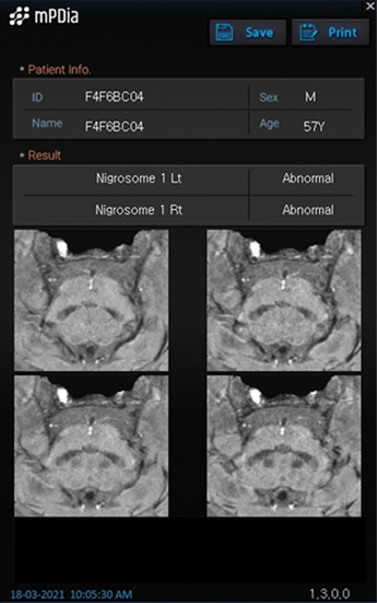
MRI-based imaging for Parkinson's Disease(PD), Nigrosome 1, can be clinically assessed and
our solutions provide information that is useful in the evaluation of patients suspected of PD.
our solutions provide information that is useful in the evaluation of patients suspected of PD.
- Acquired NET(New Excellent Technology) certification from MFDS (Ministry of Food and Drug Safety) (Cert No. 178)
- Patented in USA, Korea and Japan
- CE Marking, MFDS approved
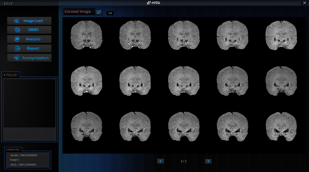
Optimal AI solution to analyze Nigrosome 1
Heuron-Parkinson automatically analyses MR images acquired via
Nigrosome 1 exposure protocol through AI algorithm, and outputs the
diagnosis of PD, assisting for medical staff to support diagnostic decision.
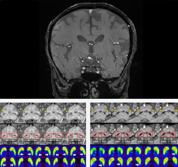
Heuron have suggested that the normal nigrosome 1 region can be identified as a hyperintense area in the dorsal aspect of the substantia nigra (SN) of healthy subjects on susceptibility map-weighted MRI (SMWI), and that its hyperintensity is lost in patients with PD.
PD diagnostic value of Nigrosome 1 in MR image was recognized academically.
Source: Automated assessment of the substantia nigra on susceptibility map-weighted imaging
using deep convolutional neural networks for diagnosis of Idiopathic Parkinson’s disease,
Parkinsonism and Related Disorders 85 (2021) 84–90 Biomarker of Parkinson's disease diagnosis
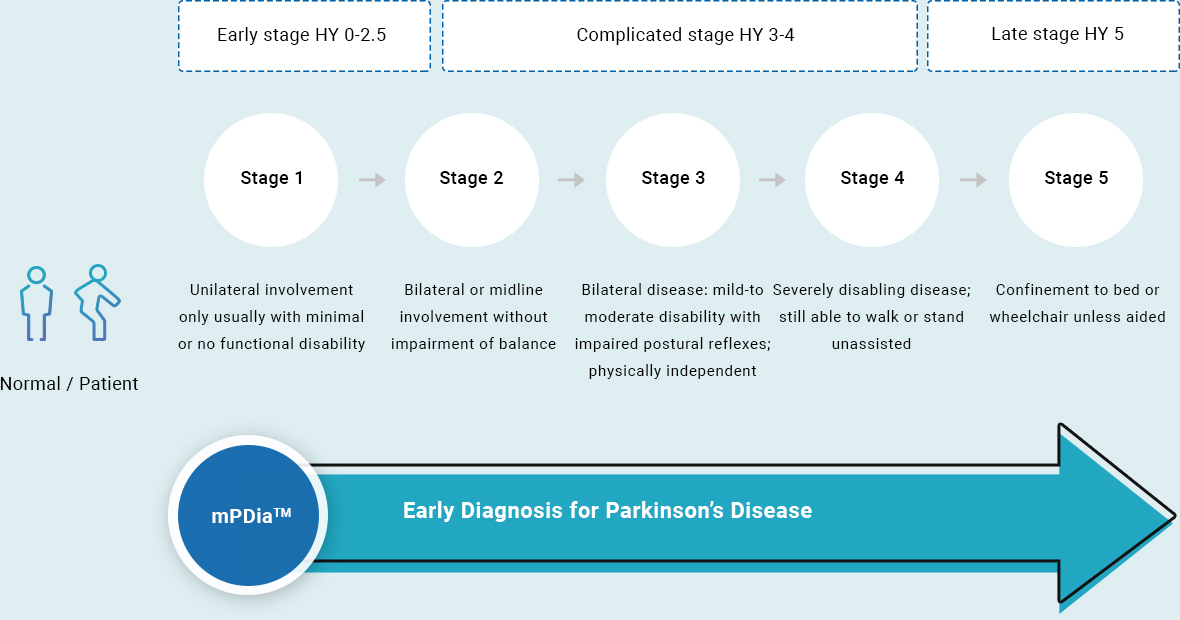
Early Diagnosis for Parkinson’s Disease
Hoehn and Yahr Scale for Staging of Parkinson’s Disease
Pre-Existing
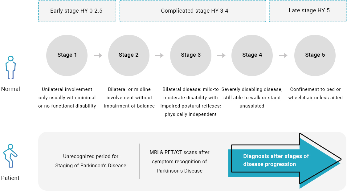
Heuron-Parkinson
Biomarker of Parkinson's Disease diagnosis
Parkinson's Disease(PD) is a progressive neurodegenerative movement disease characterized by early,
significant loss of dopaminergic neurons in the substantia nigra pars compacta(SNpc).
Nigrosome 1 is the largest subgroup of the five calbindin-poor pockets (nigrosomes) in the SNpc.
It has been reported that the maximum depletion of dopamine containing neurons in patients with PD occurs in Nigrosome 1.
DAT scan(DaT scan or Dopamine Transporter scan) will appear abnormal in which there is a loss of dopamine nerve endings,
However, DAT imaging is limited due to its lower accessibility, high cost, and radiation exposure.
Although dopaminergic functional imaging such as dopamine transporter (DAT) imaging is not essential to diagnosing PD, normal DAT imaging can exclude PD.
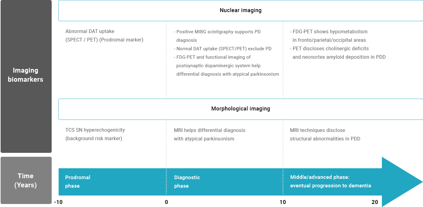
Source: Merging Clinical and Imaging Biomarkers to Tackle Parkinson’s Disease, Movement Disorders Clinical Practice, July 2017